Hip
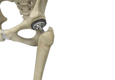
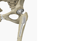
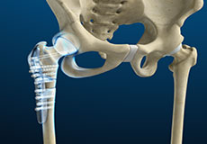
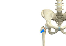
Total Hip Replacement
Total hip replacement is a surgical procedure in which the damaged cartilage and bone are removed from the hip joint and replaced with artificial components. The main indication for total hip replacement is arthritis.
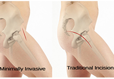
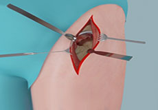
Minimally Invasive Total Hip Replacement
Minimally invasive total hip replacement is a surgical procedure performed through one or two small incisions rather than the single long incision of 10–12-inches as in the traditional approach.
Revision Hip Replacement
During total hip replacement, the damaged cartilage and bone are removed from the hip joint and replaced with artificial components. At times, hip replacement implants can wear out for various reasons and may need to be replaced with the help of a surgical procedure known as revision hip replacement surgery.
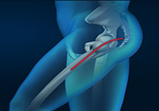
Anterior Hip Replacement
Direct anterior hip replacement is a minimally invasive hip surgery to replace the hip joint without cutting through any muscles or tendons as against traditional hip replacement that involves cutting major muscles to access the hip joint.
Posterior Hip Replacement
Posterior hip replacement is a minimally invasive surgical procedure in which the worn out or damaged surfaces of the hip joint are removed and replaced with artificial joint components made of metal, ceramic, or plastic. It is also referred to as muscle sparing surgery because key muscles and tendons are left intact while accessing the hip joint, enabling a quicker return to normal activity.
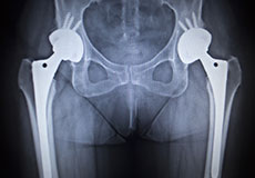
X-ray Guided Hip Replacement
Hip replacement is a surgical procedure in which the damaged hip joint is replaced with a prosthetic implant. X-ray guided hip replacement involves the use of pre-operative radiographic studies to help select the right size of the implant and to determine the proper position in which the implant should be placed.
Muscle Sparing Anterior Hip Replacement
Muscle sparing anterior hip replacement is a minimally invasive hip surgery to replace the hip joint without cutting through any muscles or tendons as compared to traditional hip replacement that involves cutting major muscles to access the hip joint.
Short-Stay and Fast-Track Hip Replacement
Short-stay and fast-track hip replacement is a minimally invasive surgical procedure in which worn-out or damaged surfaces of the hip joint are removed and replaced with prostheses. This method of treatment is known as a synchronized perioperative approach directed at decreasing surgical stress and promoting quicker postoperative recovery. The main goal of this treatment method is to reduce the necessity of hospitalization to no more than 3 days while maintaining very high patient satisfaction and few complications.

Rapid Recovery Hip Replacement
Rapid recovery hip replacement is an innovative and minimally invasive surgical procedure to replace a damaged hip joint with a prosthesis. It is associated with minimal post-operative pain and discomfort and a shorter recovery time. The surgery can often be performed as an outpatient procedure.
Hip Surgery
Hip surgery is a surgical procedure for the treatment of a hip injury or hip condition. The procedure involves repairing diseased or damaged structures of the hip joint in order to eliminate pain and restore normal hip function.
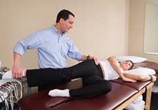
Physical Examination of the Hip
The hip joint is a ball and socket joint. The ball is the head of the femur (thighbone) that fits in the acetabulum (the hip socket). Tendons, muscles, and ligaments hold the joint in place. Articular cartilage covers the acetabulum and the femoral head. The synovial membrane of the hip joint secretes the synovial fluid which lubricates the joint enabling smooth movement.
Osteoarthritis of the Hip
Osteoarthritis, also called degenerative joint disease, is the most common form of arthritis. It occurs most often in the elderly. This disease affects the tissue covering the ends of bones in a joint called cartilage. In osteoarthritis, the cartilage becomes damaged and worn out, causing pain, swelling, stiffness and restricted movement in the affected joint. Although osteoarthritis may affect various joints including the hips, knees, hands, and spine, the hip joint is most commonly affected. Rarely, the disease may affect the shoulders, wrists, and feet.
Femoroacetabular Impingement
Femoroacetabular impingement (FAI) is a condition characterized by excessive friction in the hip joint from the presence of bony irregularities. These cause pain and decreased range of hip motion.
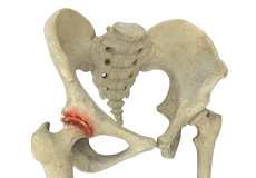
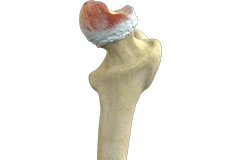
Femoral Neck Fracture
The hip is a ball-and-socket joint made up of the head of the thigh bone or femur that acts as the ball and fits into the rounded socket of the hip bone or acetabulum. The neck of the femur is the region just below the ball of the hip joint.
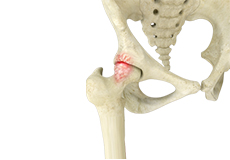
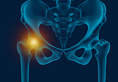
Hip Osteonecrosis
Hip osteonecrosis occurs due to disruption of the blood supply to the highest part of the thigh bone (femoral head). Due to lack of nourishment, the bone tissue of the femoral head dies and gradually collapses, which may further lead to degeneration of the underlying cartilage.
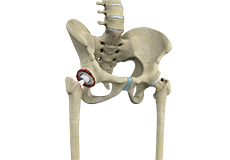
Hip Joint
The hip joint is the largest weight-bearing joint in the human body. It is also referred to as a ball and socket joint and is surrounded by muscles, ligaments, and tendons. The thigh bone or femur and the pelvis join to form the hip joint.
Any injury or disease of the hip will adversely affect the joint's range of motion and ability to bear weight.
The hip joint is made up of the following:
- Bones and joints
- Ligaments of the joint capsule
- Muscles and tendons
- Nerves and blood vessels that supply the bones and muscles of the hip
Bones and Joints
The hip joint is the junction where the hip joins the leg to the trunk of the body. It is comprised of two bones: the thigh bone or femur and the pelvis which is made up of three bones called ilium, ischium, and pubis. The ball of the hip joint is made by the femoral head while the socket is formed by the acetabulum. The Acetabulum is a deep, circular socket formed on the outer edge of the pelvis by the union of three bones: ilium, ischium, and pubis. The lower part of the ilium is attached by the pubis while the ischium is considerably behind the pubis. The stability of the hip is provided by the joint capsule or acetabulum and the muscles and ligaments which surround and support the hip joint.
The head of the femur rotates and glides within the acetabulum. A fibrocartilagenous lining called the labrum is attached to the acetabulum and further increases the depth of the socket.
The femur or thigh bone is one of the longest bones in the human body. The upper part of the thigh bone consists of the femoral head, femoral neck, and greater and lesser trochanters. The head of the femur joins the pelvis (acetabulum) to form the hip joint. Next, to the femoral neck, there are two protrusions known as greater and lesser trochanters which serve as sites of muscle attachment.
Articular cartilage is the thin, tough, flexible, and slippery surface lubricated by synovial fluid that covers the weight-bearing bones of the body. It enables smooth movements of the bones and reduces friction.
Ligaments
Ligaments are fibrous structures that connect bones to other bones. The hip joint is encircled with ligaments to provide stability to the hip by forming a dense and fibrous structure around the joint capsule. The ligaments adjoining the hip joint include:
- Iliofemoral ligament: This is a Y-shaped ligament that connects the pelvis to the femoral head at the front of the joint. It helps in limiting the over-extension of the hip.
- Pubofemoral ligament: This is a triangular shaped ligament that extends between the upper portion of the pubis and the iliofemoral ligament. It attaches the pubis to the femoral head.
- Ischiofemoral ligament: This is a group of strong fibers that arise from the ischium behind the acetabulum and merge with the fibers of the joint capsule.
- Ligamentum teres: This is a small ligament that extends from the tip of the femoral head to the acetabulum. Although it has no role in hip movement, it does have a small artery within that supplies blood to a part of the femoral head.
- Acetabular labrum: The labrum is a fibrous cartilage ring which lines the acetabular socket. It deepens the cavity, increasing the stability and strength of the hip joint.
Muscles and Tendons
A long tendon called the iliotibial band runs along the femur from the hip to the knee and serves as an attachment site for several hip muscles including the following:
- Gluteals: These are the muscles that form the buttocks. There are three muscles (gluteus minimus, gluteus maximus, and gluteus medius) that attach to the back of the pelvis and insert into the greater trochanter of the femur.
- Adductors: These muscles are located in the thigh which helps in adduction, the action of pulling the leg back towards the midline.
- Iliopsoas: This muscle is located in front of the hip joint and provides flexion. It is a deep muscle that originates from the lower back and pelvis and extends up to the inside surface of the upper part of the femur.
- Rectus femoris: This is the largest band of muscles located in front of the thigh. They also are hip flexors.
- Hamstring muscles: These begin at the bottom of the pelvis and run down the back of the thigh. Because they cross the back of the hip joint, they help in extension of the hip by pulling it backward.
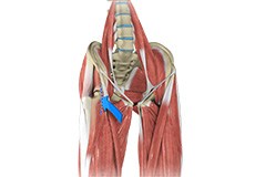
Nerves and Arteries
Nerves of the hip transfer signals from the brain to the muscles to aid in hip movement. They also carry the sensory signals such as touch, pain, and temperature back to the brain.
The main nerves in the hip region include the femoral nerve in the front of the femur and the sciatic nerve at the back. The hip is also supplied by a smaller nerve known as the obturator nerve.
In addition to these nerves, there are blood vessels that supply blood to the lower limbs. The femoral artery, one of the largest arteries in the body, arises deep in the pelvis and can be felt in front of the upper thigh.
Hip Movements
All of the anatomical parts of the hip work together to enable various hip movements. Hip movements include flexion, extension, abduction, adduction, circumduction, and hip rotation.